Chemistry 2019
40 x 64 x 3 in (h x w x d)
Honorable Mention, 2019
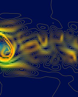
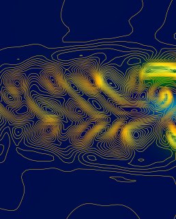
Autofluorescence microscopy can be used to visualize microscopic structures within a thin tissue section, without staining or sample modification, by exciting fluorescent chemical compounds that already exist within the tissue. Three different fluorescence filters, which show up as different colors, highlight structures of interest in a thyroid cancer tissue sample. The Eberlin lab is incorporating this technique into their molecular imaging workflow to develop a multimodal tool for improved thyroid cancer diagnosis. This image was acquired in the Microscopy and Imaging Facility at UT Austin.
Credit:
Rachel DeHoog
Chemistry Graduate Student