Nash Family Department of Pharmacological Sciences
Description:
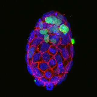
This image shows a four-day old mouse embryo. All cells of the embryo are represented in blue and cell boundaries are depicted in red. Only the cells shown in green will give rise to the whole embryo including skin, bones, fat, muscles, and the nervous system including the brain. Green-negative cells will form extra-embryonic tissues like the placenta and the yolk sac.
Bio:
Agata Kurowski, PhD, is a senior scientist with a focus on methylation-pattern-regulation during mouse development. She uses developmental as well as high-throughput methods to understand epigenetic changes. She obtained her PhD from the University of Cambridge, where she contributed to the understanding of how the pluripotency network is established in the early mouse embryo.
Research Focus:
Mechanisms of epigenetic regulation in cell differentiation and disease.