Human microglia in vivo , 2023
40 x 40 in (h x w)
100 USD
Confocal image
for sale

Genetics and Genomic Sciences
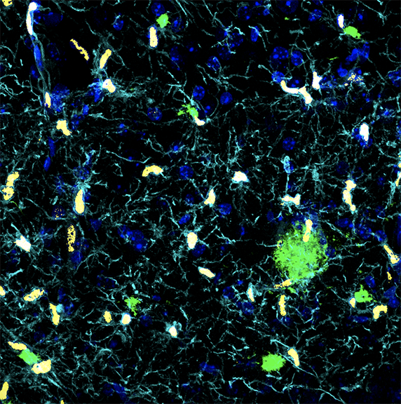
Human microglia was xenotransplanted in the brain of a mouse model of Alzheimer's Disease (AD).
This cutting-edge technique allows us to study human microglial genetics in vivo.
Confocal image:
- Cyan for microglial cells
- Yellow for a nuclear marker of human microglia
- Green for amyloid plaques (a hallmark of AD)
- Blue for nuclei of all cell types.

Printed artwork:
8” x 8” = $50
14” x 14” = $100

Mehr von The Friedman Brain Institute

Artificial Intelligence Dreams of a Cheek Cell , 2022
40 x 40 cm (h x w)
The Friedman Brain Institute
Neuronal eclipse
56 x 42 in (h x w)
The Friedman Brain Institute
YB-1 over-expression in cortical neurons , 2022
60 x 89 in (h x w)
Primary cortical neurons overexpressing YB-1, immunostained with Beta-III tubulin and imaged on a Zeiss LSM 880 confocal
The Friedman Brain Institute
Maze , 2022
50 x 66 in (h x w)
Immunoflourescence staining
The Friedman Brain Institute
La Danse , 2022
80 x 105 in (h x w)
Confocal microscopy images of (hiPSC)-derived neurons and brain organoids were taken and the figures were digitally vectorized in Adobe Illustrator to create the composition. Neurons were immunostained for MAP2, and the ventricular-like zone (neural roset
The Friedman Brain Institute