Human Deep Brain , 2023
50 x 50 in (h x w)
150 USD
for sale

Nash Family Department of Neuroscience

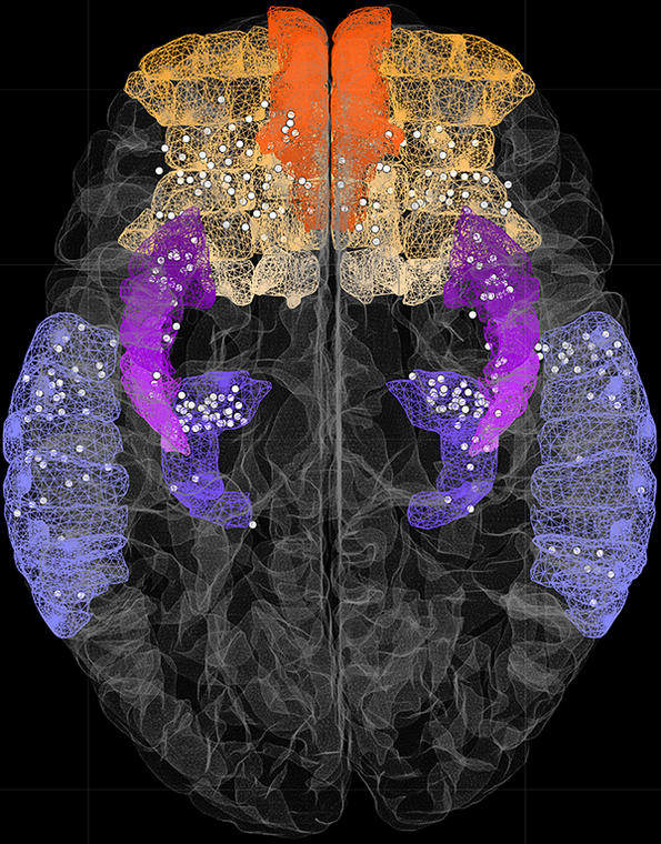
This image depicts the location of invasive stereotactic electrodes implanted deep in the brain of several intractable epilepsy patients at MSW.
Each white dot represents a single contact capable of recording brain activity.

Electrodes located in a variety of brain areas, highlighted in color, are depicted, including the orbitofrontal and ventromedial prefrontal cortices, insula, hippocampus and superior temporal sulcus.

Printed artwork:
10”(h) x 8” (w) = $100
20”(h) x 16” (w) = $150

Mehr von The Friedman Brain Institute

Artificial Intelligence Dreams of a Cheek Cell , 2022
40 x 40 cm (h x w)
The Friedman Brain Institute
Neuronal eclipse
56 x 42 in (h x w)
The Friedman Brain Institute
YB-1 over-expression in cortical neurons , 2022
60 x 89 in (h x w)
Primary cortical neurons overexpressing YB-1, immunostained with Beta-III tubulin and imaged on a Zeiss LSM 880 confocal
The Friedman Brain Institute
Maze , 2022
50 x 66 in (h x w)
Immunoflourescence staining
The Friedman Brain Institute
La Danse , 2022
80 x 105 in (h x w)
Confocal microscopy images of (hiPSC)-derived neurons and brain organoids were taken and the figures were digitally vectorized in Adobe Illustrator to create the composition. Neurons were immunostained for MAP2, and the ventricular-like zone (neural roset
The Friedman Brain Institute