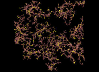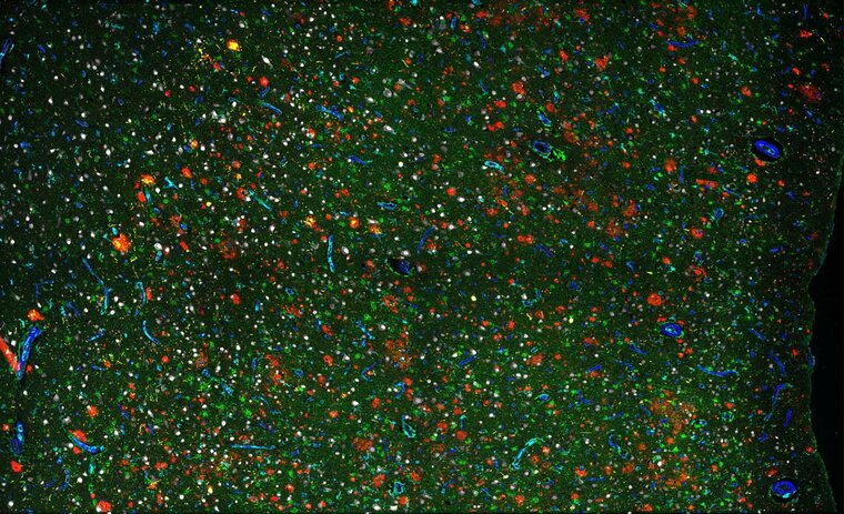
Field of poppies
, 2022
40 x 59 in (h x w)
multiplexed immunofluorescence imaging
for sale
Nash Family Department of Neuroscience
Description:
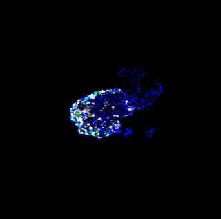
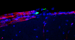
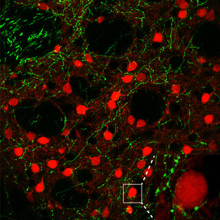
In Alzheimer’s disease, the brain shows accumulations of toxic proteins such as amyloid-β (red) and tau (yellow). Brain cell types shown here include astrocytes (green), blood vessels (blue), neurons (white), and microglia (cyan).
Bio:
The Hof lab uses classical neuropathology and modern quantitative detection to investigate neuronal susceptibility in normal aging and in neuropsychiatric disease. We also study mammalian brain evolution, particularly in cetaceans and great apes.
Research Focus: Aging and Alzheimer's disease
Funding:
Research for which this image was supported by the National Institute On Aging of the National Institutes of Health under Award Number 3R01AG059028-01S2.