Tree of Life , 2022
50 x 47 in (h x w)
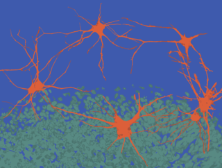
Confocal image of viral dTomato expressed in cerebellar Purkinje neuron, rainbow pseudo-coloring
for sale

Nash Family Department of Neuroscience

Description:
Fluorescent labeling of highly elaborate branching structure of a Purkinje neuron in the cerebellum

Bio:
Dr. Molly Heyer is an instructor in the lab of Paul Kenny at Mount Sinai. She studies role of regulatory small RNAs (microRNAs) in cerebellar neuron function and dysfunction related to psychiatric disorders including schizophrenia and autism spectrum disorder.

Research focus: Schizophrenia and autism spectrum disorder

More from The Friedman Brain Institute

Artificial Intelligence Dreams of a Cheek Cell , 2022
40 x 40 cm (h x w)
The Friedman Brain Institute
Neuronal eclipse
56 x 42 in (h x w)
The Friedman Brain Institute
YB-1 over-expression in cortical neurons , 2022
60 x 89 in (h x w)
Primary cortical neurons overexpressing YB-1, immunostained with Beta-III tubulin and imaged on a Zeiss LSM 880 confocal
The Friedman Brain Institute
Maze , 2022
50 x 66 in (h x w)
Immunoflourescence staining
The Friedman Brain Institute
La Danse , 2022
80 x 105 in (h x w)
Confocal microscopy images of (hiPSC)-derived neurons and brain organoids were taken and the figures were digitally vectorized in Adobe Illustrator to create the composition. Neurons were immunostained for MAP2, and the ventricular-like zone (neural roset
The Friedman Brain Institute