First line of defense
, 2022
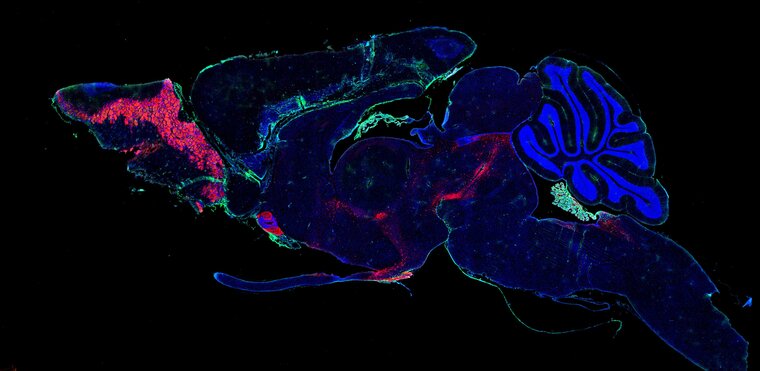
Immunofluorescence staining of fixed frozen brain section & widefield slide-scanning microscopy
not for sale
Nash Family Department of Neuroscience
Description:
The image shows a sagittal brain section of a mouse that has been injected with a viral mimetic to investigate the anti-viral response in the brain. The blue stain shows cell nuclei, the red stain shows dopamine producing cells, and the green strain shows cells that produce Mx1, an anti-viral response gene.
Bio:
Yajing Xu is a postdoctoral researcher in the Department of Neuroscience where she investigates microglial response to peripheral infections. She received her PhD from UCL in London, where she studied the role of microglia in the postnatal development of spinal cord pain circuitry.
Research focus: Microglia & brain response to peripheral immune challenge