Department of Genetics and Genomics Sciences
Description:
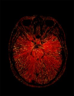
Immune cells in the brain fighting against Alzheimer's Disease pathology
My artwork was created using immunofluorescence technique. Mouse brain tissue was section on a cryostat, then free-floting stained with antibody Iba1 to mark microglial cells. Image was acquired in a confocal microscope, with a 40X magnification, using Z-stack projection. Image was processed and modified using Image J software.
Bio:
Carmen Romero-Molina is a Postdoc at Dr. Goate's Lab (Genetic and Genomics Dept), investigating the role of microglial cells in Alzheimer's Disease. She got her PhD in Neuroscience by University of Seville (Spain), also performing short-period rotations at Imperial College of London (London, UK) and Harvard University (Boston, US).
Research Focus: The role of microglial cells in Alzheimer's Disease