Department of Psychiatry
Description:
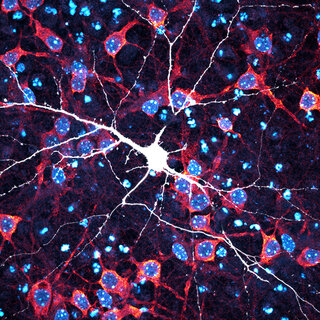
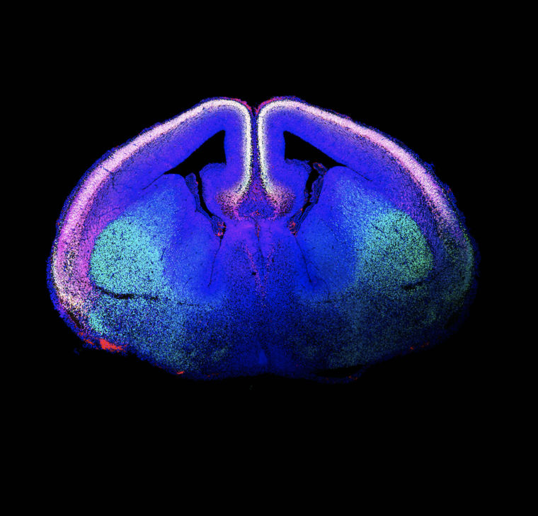
This image shows us in white/yellow the first neurons that are born in the embryonic mouse cortex. These neurons were born in the ventricular zone (zone in the cortex closer to the ventricles) and have traveled to their ultimate position in the cortical plate. From now on, neurons will continue to be born and travel to their ultimate position on top of these first neurons, until the functional cortex is formed.
Bio:
Marta Garcia-Forn join the De Rubeis lab in January 2020 after a PhD in Biomedicine at the University of Barcelona in Esther Perez-Navarro's lab. Martha studies the development and connectivity of the cortex of mice modeling DDX3X syndrome, and their relationship to behavioral deficits. Martha is supported by the Seaver Foundation and Fundacion Alfonso Martin Escudero.
The De Rubeis lab is part of the Seaver Autism Center for Research and Treatment and the Psychiatry department at ISMMS. The aim of the lab is to understand the cellular and molecular mechanisms underlying DDX3X syndrome, a rare genetic condition associated with intellectual disability and autism spectrum disorder manifesting primarily in girls.