La Danse
, 2022
80 x 105 in (h x w)
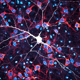
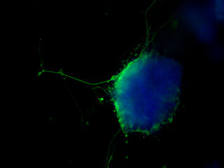
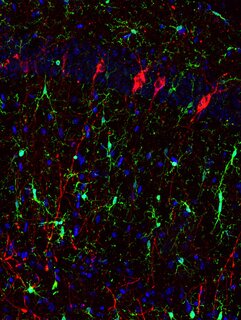
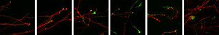
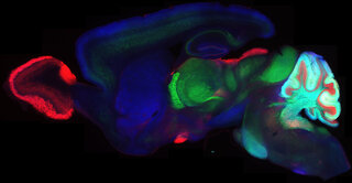
Confocal microscopy images of (hiPSC)-derived neurons and brain organoids were taken and the figures were digitally vectorized in Adobe Illustrator to create the composition. Neurons were immunostained for MAP2, and the ventricular-like zone (neural roset
for sale
Nash Family Department of Neuroscience
Description:
Neurons dance together atop a field of cell nuclei in this reimagining of Henri Matisse’s 1910 painting, La Danse. A version of Matisse's painting hangs in New York City’s Museum of Modern Art.
I used images/cell cultures from my rotation in Paul Slesinger's lab - the cell lines were from human donors with high and low Polygenic Risk Scores for Alcohol Use Disorder.
I would like to credit Isabel Gameiro-Ros, PhD, who grew the neurons that went into the confocal images.
Research focus: Induced pluripotent stem cell modeling of neuropsychiatric disorders.