The Beginning of Life
, 2023
70 x 70 in (h x w)
not for sale
Department of Pharmacological Sciences
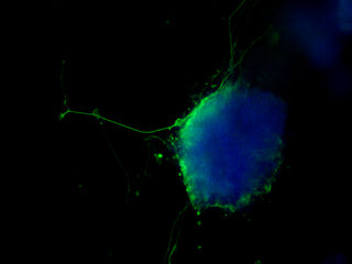
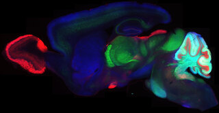
This image shows a three-day-old mouse embryo stained with antibodies against proteins that are specific to distinct cell populations.
As development proceeds, the cells shown in red (stained using an antibody against NANOG) will uniquely form the embryo including skin, bones, fat, muscles, and the nervous system including the brain.
The green cells (antibody against CDX2) will form the placenta. E-cadherin is a protein in the cell boundaries and is immuno-stained and shown in blue.
This artwork is a collection of different compositions of the two mutually exclusive cell populations and their cell boundaries.