Human Microglia in vivo #2! , 2023
50 x 50 in (h x w)
for sale

Department of Genetics and Genomic Sciences

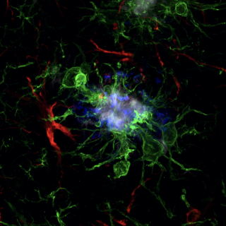
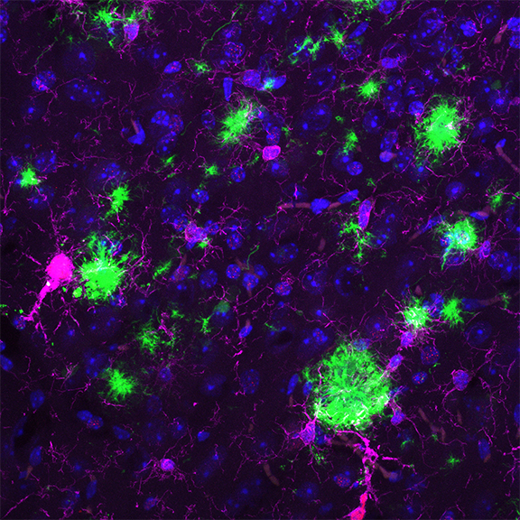
This image focuses on human microglia that were xenotransplanted in the brain of a mouse model of Alzheimers Disease.

This display can be seen very clearly and studied through our images taken on the confocal!

This confocal image consists of:

- Magenta for microglial cells
- Red for a nuclear marker of human microglia
- Green for amyloid plaques
- Blue for nuclei of all cell types

More from The Friedman Brain Institute

The Great Escape: Mapping the path to Freedom , 2024
40 x 71 in (h x w)
Photography
The Friedman Brain Institute
Neuroinflammation in a progressive supranuclear palsy brain , 2024
28 x 40 in (h x w)
Photography
The Friedman Brain Institute
Neuroinflammation in a progressive supranuclear palsy brain , 2024
28 x 40 in (h x w)
Photography
The Friedman Brain Institute
Safa Srours Brain Aneurysm , 2024
25 x 20 in (h x w)
Photography
The Friedman Brain Institute
Snowball makers , 2024
25 x 25 in (h x w)
Photography
The Friedman Brain Institute