Nash Family Department of Neuroscience
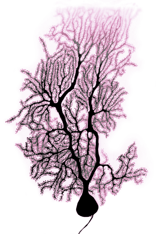
The elaborate branches and spines of a cerebellar Purkinje neuron call to mind a springtime cherry tree in bloom. This inverted and pseudocolored confocal image was obtained from an adult mouse Purkinje neuron iontophoretically filled with Lucifer Yellow fluorescent dye.
Dr. Heyer studies the roles of small RNAs in regulating Purkinje neuron function and downstream behaviors. Cerebellar abnormalities such as altered Purkinje neuron activity have been strongly linked to disorders such as autism and schizophrenia.
The Drug Discovery Institute is actively pursuing novel ways to modulate Purkinje neuron activity, which may lead to new strategies for treating these disorders.
Printed artwork:
12”(h) x 8” (w) = $100
20”(h) x 14” (w) = $150