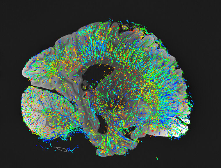
Human Brain Angio PRH
, 2023
60 x 60 in (h x w)
not for sale
Nash Family Department of Neuroscience
This image is the result of a collaboration between Drs. Bruce Fischl (Harvard, MGH), David Boas (Boston University), Irene Costantini and Francesco Pavone (LENS, Florence, Italy), and Patrick R. Hof, MD, FAAA.
Here is a single-level 7 T MRI of a human brain on which a dataset of angioarchitecture was obtained in full 3D through which the same whole hemisphere using polarization-sensitive optical coherence tomography was re-instantiated.