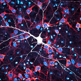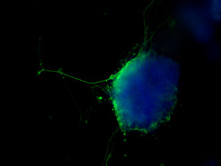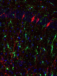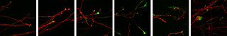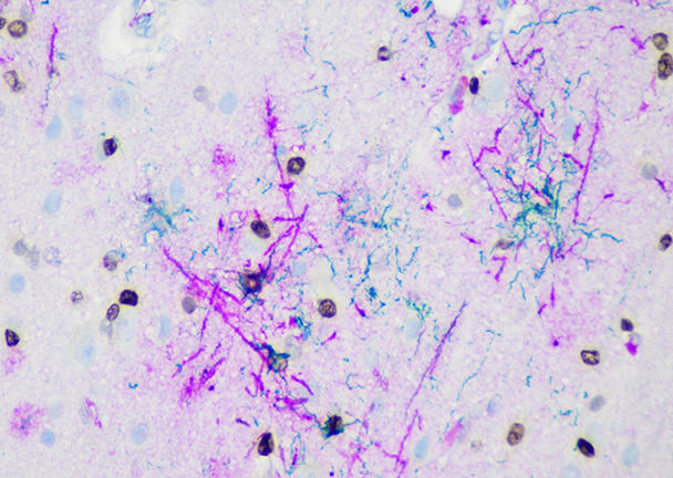
Neuroinflammation in a progressive supranuclear palsy brain
, 2024
28 x 40 in (h x w)
Photography
Department of Pathology
Immunohistochemical images of complement (C4A, purple), hyperphosphorylated tau (AT8, green), and Oligodendrocyte marker (OLIG2, brown) in frontal cortex of human postmortem progressive supranuclear palsy (PSP) and control brain tissue. When the complement and hyperphosphorylated tau overlap it appears as blue.