Rear Window
16 x 16 x 2 in (h x w x d)
for sale

Nash Family Department of Neuroscience

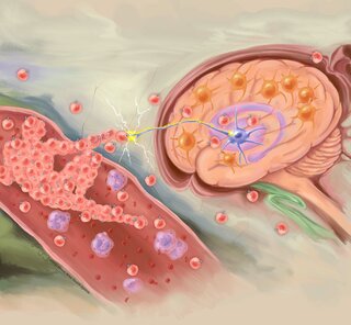
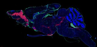
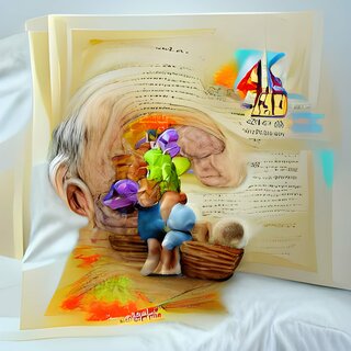
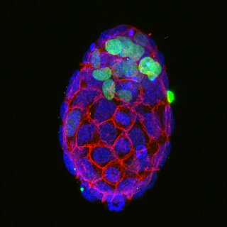
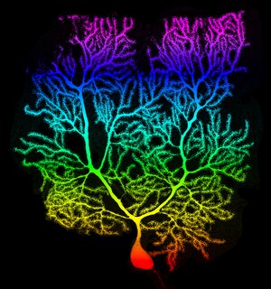
Stained proteins light up disease hallmarks (plaques and tangles) and stress markers, near or inside cells in the brain of a person with Alzheimer’s disease. These microscope images provide a window that reveals players involved in the death of brain cells in Alzheimer’s.

Research underlying this image was supported by the National Institute On Aging of the National Institutes of health under Award Number 3R01AG059028-01S2. The content is solely the responsibility of the authors and does not necessarily represent the official views of the National Institutes of Health.

More from The Friedman Brain Institute

Birth of Movement Disorder , 2022
42 x 44 in (h x w)
The Friedman Brain Institute
First line of defense , 2022
Immunofluorescence staining of fixed frozen brain section & widefield slide-scanning microscopy
The Friedman Brain Institute
Fading Memories , 2022
Adobe Photoshop
The Friedman Brain Institute
The Awakening , 2022
50 x 50 in (h x w)
Confocal microscopy imaging of an immuno-fluorescence staining of a dissected mouse embryo.
The Friedman Brain Institute
Tree of Life , 2022
50 x 47 in (h x w)
Confocal image of viral dTomato expressed in cerebellar Purkinje neuron, rainbow pseudo-coloring
The Friedman Brain Institute