Nash Family Department of Neuroscience
Description:
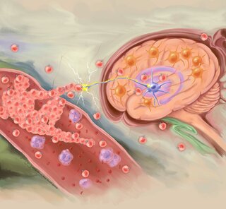
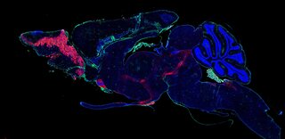
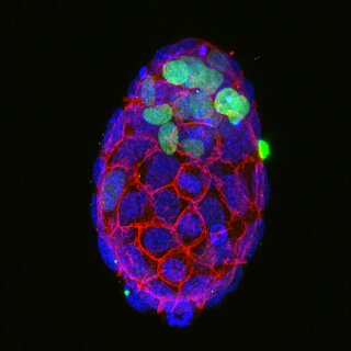
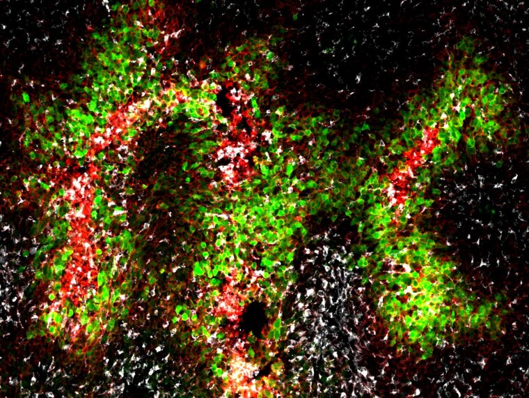
Three hypoxic zones in a brain cancer. Hypoxic tumor cells (green) surround immune cells (red and gray)
Immunoflourescence staining was used to create the artwork. Antibodies that bind to cell type specific markers were conjugated with flourescent dyes to generate the image. Additionally, tumor cells were genetically engineered to express flourescent proteins.
Bio:
I am a postdoctoral fellow at Jenny Zou and Roland Friedel laboratories. I have a PhD in Cancer Biology from Wake Forest University and was a visiting PhD scholar at Columbia University before coming to Mount Sinai. I have been researching glioblastoma biology and working to develop treatments over the past 10 years.
Research focus:
My research focus is Glioblastoma, which is the most common and lethal brain cancer.