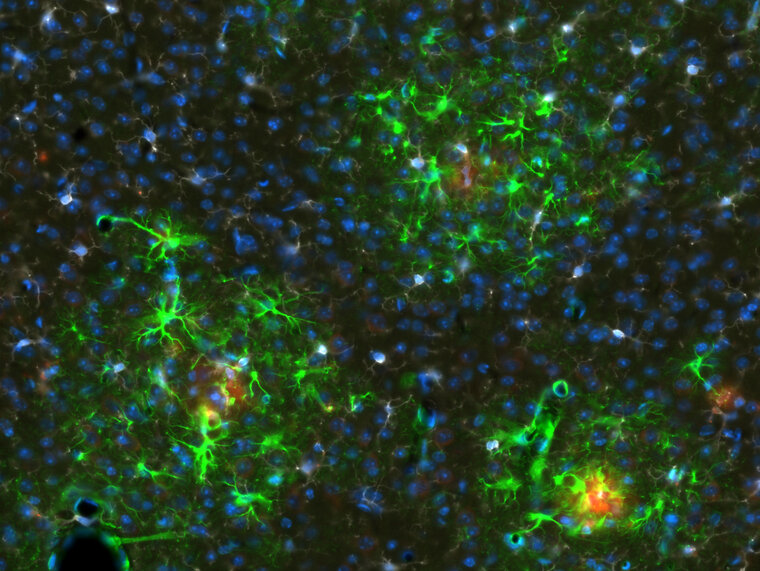
Reactive Astrocytes Surrounding Amyloid Plaque
14 x 19 in (h x w)
for sale

Nash Family Department of Neuroscience

In the cortex of 6-month Alzheimer’s Disease mouse model (APP/PS1), reactive astrocytes (immunostaining by astrocyte marker GFAP antibody, green fluorescence) surround amyloid beta (Aβ) plaque (immunostaining by Aβ antibody, Texasred fluorescence) which is pathological marker of Alzheimer’s Disease. DAPI staining (blue fluorescence) is used for nuclei of all cells.

Plus de The Friedman Brain Institute

Artificial Intelligence Dreams of a Cheek Cell , 2022
40 x 40 cm (h x w)
The Friedman Brain Institute
Neuronal eclipse
56 x 42 in (h x w)
The Friedman Brain Institute
YB-1 over-expression in cortical neurons , 2022
60 x 89 in (h x w)
Primary cortical neurons overexpressing YB-1, immunostained with Beta-III tubulin and imaged on a Zeiss LSM 880 confocal
The Friedman Brain Institute
Maze , 2022
50 x 66 in (h x w)
Immunoflourescence staining
The Friedman Brain Institute
La Danse , 2022
80 x 105 in (h x w)
Confocal microscopy images of (hiPSC)-derived neurons and brain organoids were taken and the figures were digitally vectorized in Adobe Illustrator to create the composition. Neurons were immunostained for MAP2, and the ventricular-like zone (neural roset
The Friedman Brain Institute