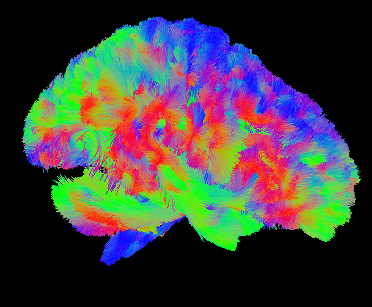
DTI Representation of Whole Brain White Matter Tracts
16 x 20 in (h x w)

Biomedical Engineering and Imaging Institute

A 3D representation of the human brain's white matter connections in the side view. These tracts connect different regions of the brain with one another, with the different colors representing their direction (blue is up/down, green is front to back, and red is across the sides of the head). The data used to generate this image was taken using MRI.

Plus de The Friedman Brain Institute

Artificial Intelligence Dreams of a Cheek Cell , 2022
40 x 40 cm (h x w)
The Friedman Brain Institute
Neuronal eclipse
56 x 42 in (h x w)
The Friedman Brain Institute
YB-1 over-expression in cortical neurons , 2022
60 x 89 in (h x w)
Primary cortical neurons overexpressing YB-1, immunostained with Beta-III tubulin and imaged on a Zeiss LSM 880 confocal
The Friedman Brain Institute
Maze , 2022
50 x 66 in (h x w)
Immunoflourescence staining
The Friedman Brain Institute
La Danse , 2022
80 x 105 in (h x w)
Confocal microscopy images of (hiPSC)-derived neurons and brain organoids were taken and the figures were digitally vectorized in Adobe Illustrator to create the composition. Neurons were immunostained for MAP2, and the ventricular-like zone (neural roset
The Friedman Brain Institute