Nash Family Department of Neuroscience
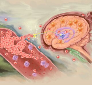
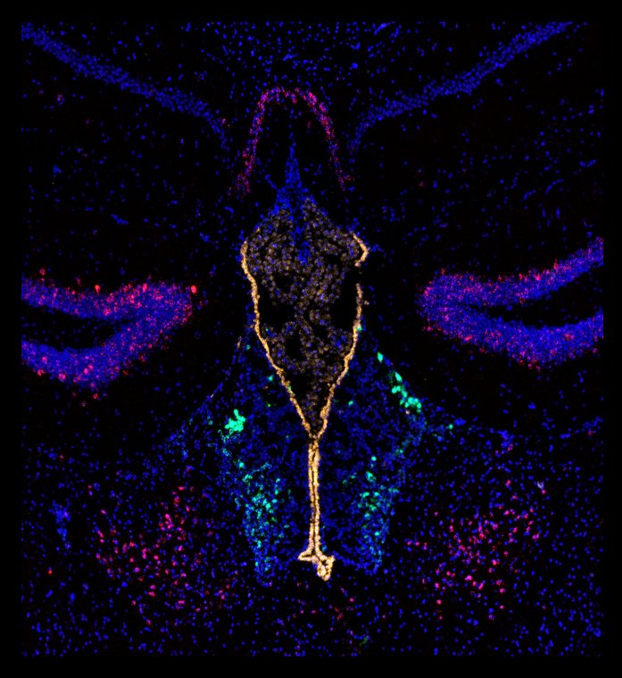
Lateral habenula (LHb) neurons, along with various cell types, exhibit responsiveness to stress, making their neuromodulation a promising avenue for the development of innovative therapies targeting major depressive disorder and substance use disorders. To gain a deeper understanding of the intricate cellular composition of the LHb, scientists employ advanced imaging techniques with high spatial resolution.
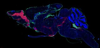
This particular image showcases a coronal section of the habenula in a mouse brain, employing the in situ hybridization technique (RNAscope). The color-coded labeling provides insight into distinct cell populations:
Cells expressing the dopamine 1 (drd1) receptor are highlighted in red.
Cells expressing tyrosine hydroxylase (TH) are depicted in green.
Cells expressing Forkhead box protein J1 (foxj1) are represented in orange.
DAPI staining in blue serves to label cell nuclei, offering a comprehensive view of the cellular architecture.
This visually captivating representation not only enhances our comprehension of the LHb's cellular diversity but also serves as a valuable tool in unraveling the intricate neurobiological mechanisms associated with stress, laying some groundwork for potential breakthroughs in treating mental health disorders.